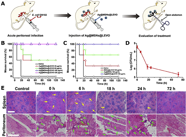
Figure 13. In vivo Evaluation of E. coli Infected Mice Peritoneal Wound Healing Effect of Ag@MSNs@LEVO.
(A) Scheme of in vivo infection and treatment procedure.
(B, C) Mice survival rates after acute peritoneal infection and treatment with Ag@MSNs@LEVO and control groups.
(D) Bacterial counts within the peritoneal cavity of mice after treatment with 160 μg/mL Ag@MSNs@LEVO.
(E) H&E staining of histological sections including spleen and peritoneum of mice after acute peritoneal infection and treatment with 160 μg/mL Ag@MSNs@LEVO. Extended lymphoid nodules on spleen and inflammatory cells on peritoneum are respectively marked with yellow and green arrows.
(A-E) Reprinted from Ref. 214, Copyright (2016), with permission from Elsevier.

