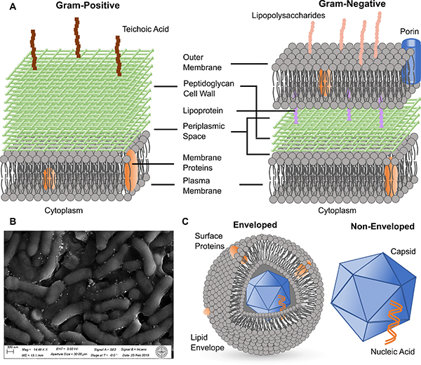
Figure 2. Microbial Pathogens.
(A) Scheme of the cell wall and membrane structures of Gram-positive (left) and Gram-negative (right) bacteria.
(B) SEM micrograph of a biofilm formed from Arthrobacter sp. mixed with Ag NPs.
(C) Scheme of the structures of enveloped (left) and non-enveloped (right) viruses.

