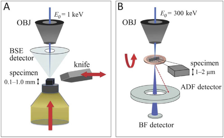
Figure 1.
(A) In SBF-SEM, a heavy-atom stained, resin-embedded biological specimen is first raster-scanned with a low energy electron beam (keV). A back scattered detector (BSD) is used to image the electrons back-scattered from a certain depth of the specimen. A diamond knife, mounted in situ, then slices off a thin layer and exposes a new surface to be imaged. The block is then raised a desired height, which dictates the cutting slice thickness and the pattern repeats. A series of consecutive images produces a volumetric dataset. (B) In STEM, 300 keV incident electrons have multiple ways of interacting with a thick (>1 μm) resin-embedded material: a red dotted line represents electrons that arrive at the bottom of the specimen with large displacement from the center, which land on the high angle-annular dark field (HAADF) detector; blue line represents the electrons along the central axis of the specimen which land on the on-axis bright field (BF) detector. When imaging thick specimens, the use of HAADF detector produces blurry unfocused images, while BF detector’s larger depth-of-field allows imaging of thick specimens without the loss of focus through the entire thickness of the plastic section. BF combined with tomography allows one to obtain three-dimensional structural information.

