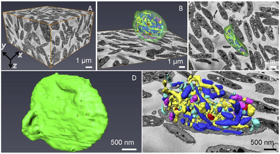
Figure 2.
SBF-SEM acquired 3D data of human resting platelets. (A) xy, yz and xz planes (orthoslices) define a volume of 7.5 μm in z, containing more than one hundred platelets. Each voxel is 6.8 nm x 6.8 nm x 30 nm. A complete platelet surface rendering in xz (B) and xy (C) views, together with an xy orthoslice, which shows organization of different membrane-bound organelles: open canalicular system (yellow), closed canalicular system (cyan), mitochondria (purple), alpha granules (blue), dense granules (red), dense cores (burgundy), plasma membrane (green). (D) Segmented outer plasma membrane of a complete platelet. (E) Magnified segmented platelet organelles without the membrane together with the xy orthoslice.

