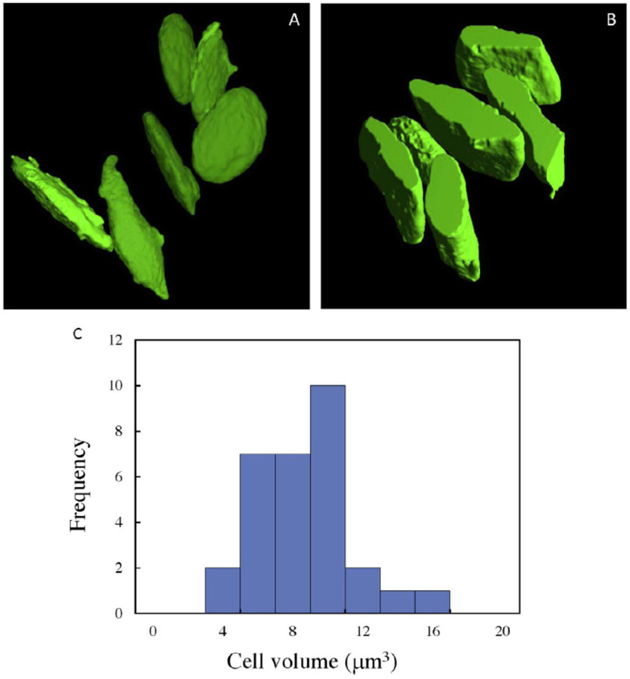
Figure 4.
Surface rendered plasma membrane of six unactivated platelets obtained by (A) SBF-SEM, and (B) STEM tomography reveals the characteristic platelet shape. In (B) only about half of the platelets are contained within the 1.5 μm-thickness of the plastic section. (C) Histogram of platelet volumes from 30 cells, obtained by SBF-SEM, with mean value of 7.7 μm3 ± 2.6 μm3 (± std. dev.).

