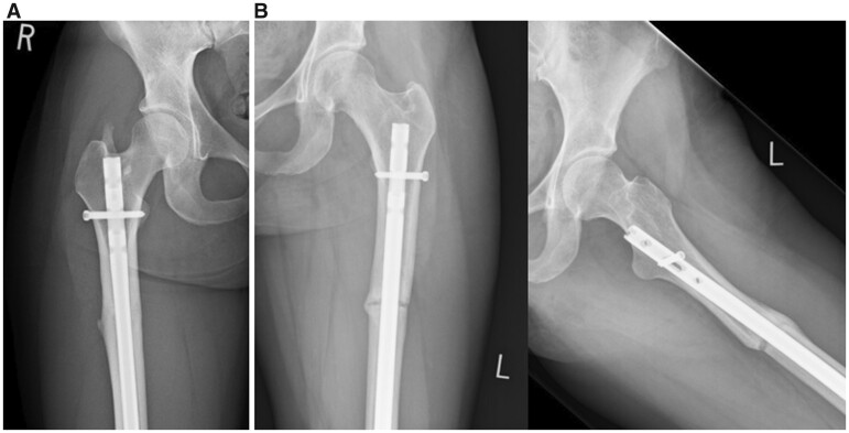
Fig. 7.
Post-operative X-ray images demonstrating (A) AP image of a patient with HO at the proximal nail insertion. (B) AP and lateral images of a delayed union at 6 months. The osteotomy had not been actively compressed during surgery in this particular case. (i) 19 × 32 mm (300 × 300 DPI) and (ii) 47 × 32 mm (300 × 300 DPI).

