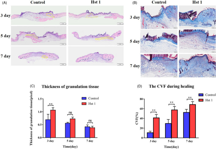
FIGURE 2.

Distribution of granulation tissue and extracellular matrix during healing. A, The HE staining showed the thickness of granulation tissue. B, Statistical data of granulation tissue area during healing process. Staining of interstitial components in granulation tissue. C, Masson staining was used to stain the collagen fibres in the skin sections during the healing process. D, Collagen volume fraction (CVF) during healing process, Vertical axis, CVF; horizontal axis, time. Data are shown as mean ± SE. (*P < .05; **P < .01)
