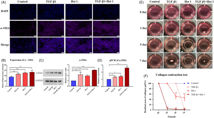
FIGURE 6.

Transformation ability to myofibroblast and function of 3T3 cells. A‐D, The control group was cultured with DMEM medium (10% FBS), and the experimental group was added with 10 ng/mL TGF ‐β1, 10 μmol/L Hst 1 or both of them, respectively. Immunofluorescence staining (DAPI was labelled blue fluorescence and α‐SMA was purple), western blot and quantitative PCR were used to detect the expression of α‐SMA. E, Collagen contractive function of fibroblasts. Fibroblasts were added into the collagen of rat tail after gelatinization, and the factors were added to stimulate the cells as mentioned above. F, The contraction area of collagen was calculated by the image analyser. The data of each group were compared with the control group. The data provided are one of the representative data. All experiments were repeated three times in an independent occasion. Data are shown as mean ± SE. (*P < .05; **P < .01)
