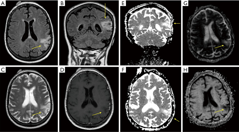
Figure 1.
Images from brain MRI at diagnosis. Arrows point at a triangular focal alteration of the cortical-subcortical signal that can be observed on the left parietal lobe, associated to a slimming of the cortex and a mild ampliation of the adjacent subarachnoid space, hyperintense in FLAIR (A,B) and T2 (C) sequences, and hypointense in T1 sequence (D). The diffusion-weighted magnetic resonance imaging (DWI) technique shows a cortical restricted diffusion in this same area (E,F). Correlation with apparent diffusion coefficient (ADC) and exponential apparent diffusion coefficient (eADC) maps is shown in (G) and (H) respectively.

