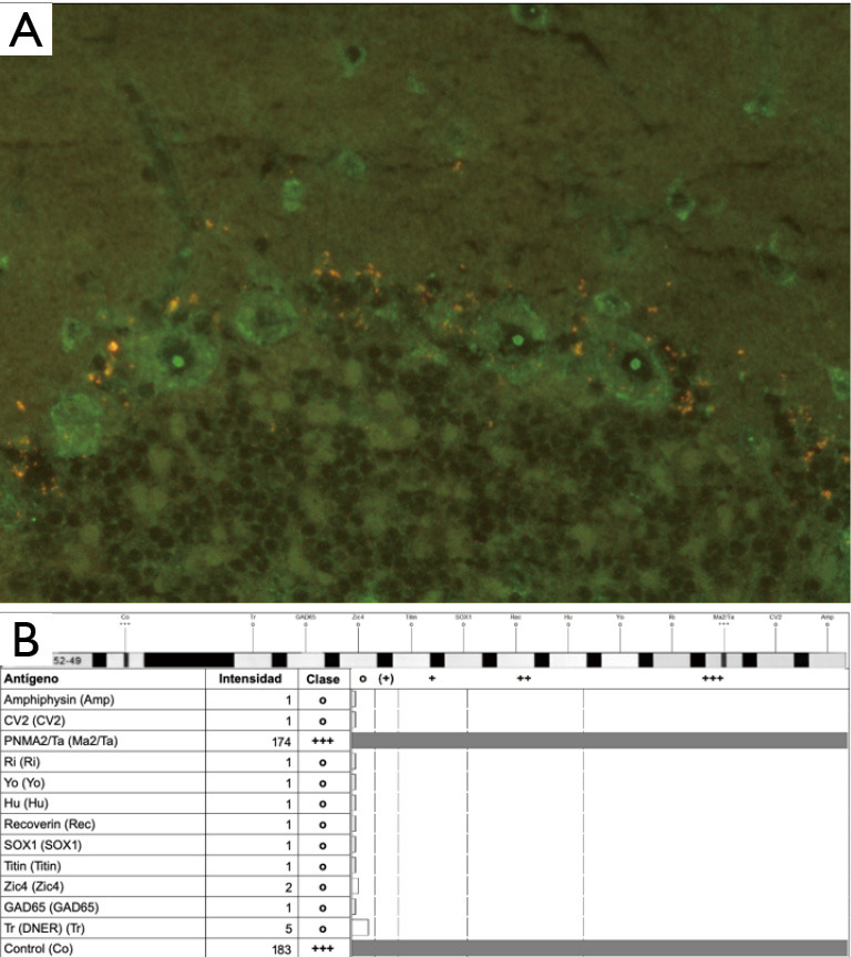
Figure 2.

Detection of anti-Ma2 antibodies. (A) Indirect immunofluorescence demonstrating presence of anti-Ma2 Abs from the peripheral blood sample of our patient. Anti-Ma2 Abs are shown in cerebellar dentate nucleus Purkinje cells, with a high uptake in the neuronal nucleolus. (Magnification times: 40×/0.75). (B) Western blot analysis revealing a high intensity band corresponding to the anti-Ma2 antibody, with a quantitative value of expression of 95 UA/100.
