Figure 6.
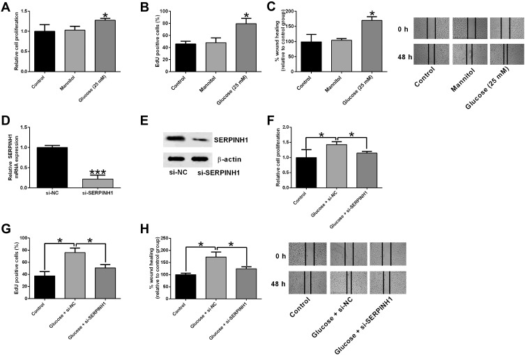
SERPINH1 silence attenuated the high glucose-induced increase in cell proliferation and migration of HRECs. (A) The cell proliferation of HRECs after different treatments (control, mannitol, 25 mM glucose) was determined by CCK-8 assay. (B) EdU assay was used to determined HREC proliferation after different treatments (control, mannitol, 25 mM glucose). (C) The HREC migration after different treatments (control, mannitol, 25 mM glucose) was determined by wound healing assay. (D) The mRNA and (E) protein expression levels of SERPINH1 in HRECs after being transfected with si-NC or si-SERPINH1 were determined by qRT-PCR and Western blot assay, respectively. (F–H) HRECs were transfected with si-NC or si-SERPINH1 followed by treating with 25 mM glucose, the cell proliferation and migration of HRECs were determined by CCK-8, EdU and wound healing assay, respectively. *p<0.05.

