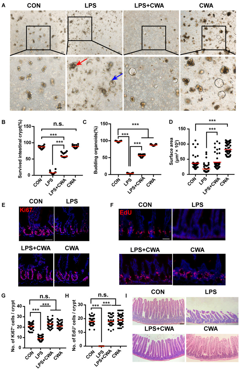
FIGURE 4.
CWA preserves ISCs viability and promotes epithelial regeneration. (A) Representative bright-field images of crypts after 4 days of culture in ENR medium. Red arrowhead indicates collapsed organoids, blue arrowhead indicates survived organoids. Scale bars, 100 μm. (B) Survival rate of crypts isolated from each group. (C) Organoid budding, percentage of total organoids per well. (D) Size of SI organoids cultured in ENR medium. (E) Representative confocal microscopy images of Ki67 positive cells in crypts. Scale bars, 50 μm. (F) Representative immunofluorescence images of EdU positive cells. Scale bars, 50 μm. (G) Quantification of Ki67+ cells in each crypt. (H) Quantification of EdU+ cells in each crypt. (I) H&E staining of jejunum sections at 24 h after CWA administration. Scale bars, 100 μm. ***P < 0.001 by two-sided, unpaired t-test. All data represent at least three independent experiments.

