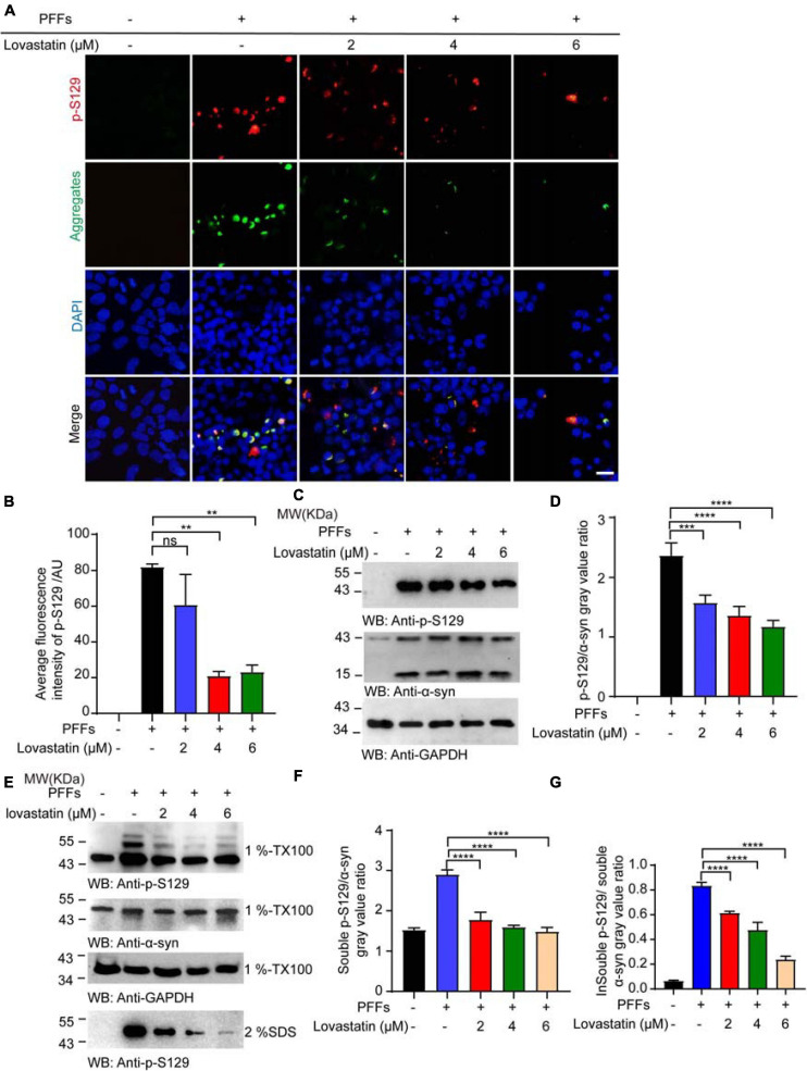
FIGURE 2.
Lovastatin attenuates α-syn phosphorylation in GFP-α-syn-HEK293 cells. (A) Immunofluorescence showing that p-S129 co-localized with α-syn aggregates. Intracellular p-S129 α-syn (red) was decreased in the lovastatin group. (B) Quantitative analysis of p-S129 α-syn fluorescence. (C) Western blot analysis of the p-S129 level. (D) Quantification of the p-S129 level in Western blot. Results are normalized to total α-syn. (E) Western blot analysis of soluble and insoluble p-S129 α-syn. (F) Quantification of soluble p-S129 α-syn. Results are normalized to total α-syn. (G) Quantification of insoluble p-S129 α-syn. Results are normalized to total α-syn. All data are means ± SEM, **P < 0.01, ***P < 0.001, and ****P < 0.0001, ns: not statistically significant, one-way ANOVA with Tukey’s multiple comparisons test. Bar = 20 μm. All experiments were performed in triplicate for at least three independent times.

