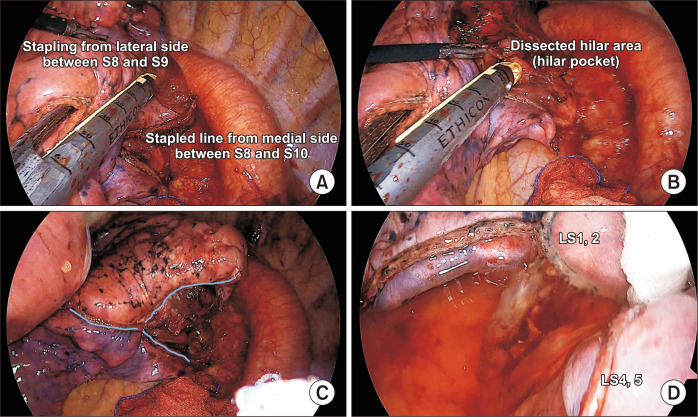
Fig. 5.
(A) Peripheral stapling at different angles when starting the division of the intersegmental plane. (B) Placement of the stapler tip into the hilar pocket after further dissection of the hilar area. (C) Triangular stapled line after left 9,10 segmentectomy. (D) A U-shaped (or V-shaped) stapled line after left 3 segmentectomy

