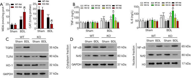
Figure 3.
TGR5−/− BDL mice showed increased oxidative stress and pro-inflammatory response. WT or TGR5−/− mice underwent BDL. (A) MDA and CAT activities in liver tissues on Day 10 (n=3 per group); (B) TNF-α and IL-6 level on Day 1, 3, 7, and 10 measured by ELISA (n=3 per group); (C) TGR5, TLR4, and HO-1 expression in liver on Day 10 (n=3 per group); (D) the expression levels of NF-κB (nuclear and cytoplasmic) and Nrf2 (nuclear and cytoplasmic) in liver on day 10 (n=3 per group). **P<0.01, ***P<0.001 (vs. control); #P<0.05, ##P<0.01 (vs. WT). BDL, bile duct ligation. MDA, malondialdehyde; CAT, catalase.

