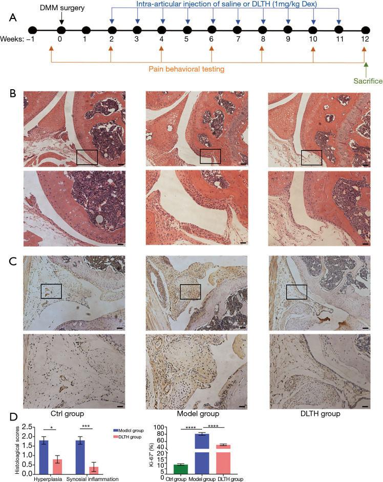
Figure 2.
H&E and immunohistochemical staining of DLTH treatment in DMM-induced osteoarthritis. (A) Schematic of the experimental study design. Three groups of male C57BL/6J mice were studied: Ctrl group, Model group and DLTH group. Mice in the Model and DLTH groups received DMM surgery on the right hind knees at 10 weeks of age, while mice in the Ctrl group underwent sham operations in the same knee. Mice in the DLTH group were subjected to joint injection of 10 µL DLTH (1 mg/kg Dex per mouse) into the right knee once a week from week 2 to week 11, while mice in the Ctrl and Model groups were given an equal volume of saline in the same knee. The timelines for DMM surgery, IA-DLTH injection, and behavioral pain testing are shown. (B) Histological analyses of synovial joints in DLTH-treated OA mice. Representative photographs of mice knee joints stained with H&E were displayed. Scale bar, 200 µm (upper panel) and 100 µm (lower panel). Black boxes represented the magnification area in the corresponding figure below. (C) Ki-67 staining of synovia in DLTH-treated OA mice. Representative photographs of mice knee joints stained with Ki-67 are shown. Scale bar, 200 µm (upper panel) and 100 µm (lower panel). Black boxes represented the magnification area in the corresponding figure below. (D) Quantification of hyperplasia and synovial inflammation using semiquantitative histology scoring system; Ki-67 positive cells in the synovia are displayed. All data were expressed as mean ± SEM. *P<0.05; ***P<0.001; ****P<0.0001. Ctrl, control; DLTH, dexamethasone-loaded thermo-sensitive hydrogel; DMM, destabilization of the medial meniscus; H&E, hematoxylin and eosin; IA, intra-articular; OA, osteoarthritis; SEM, standard error of the mean.

