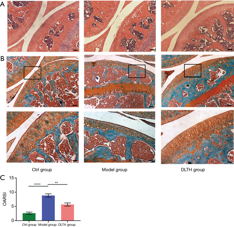
Figure 5.
S&F and H&E staining of DLTH treatment in DMM-induced osteoarthritis. (A,B) H&E and S&F staining of knee joint medial compartment cartilage and subchondral bone in DMM-induced mice. Scale bar: 200 µm (upper panel) and 100 µm (lower panel). Black boxes represented the magnification area in the corresponding figure below. (C) Quantitative analyses of OARSI scores of all groups. All data were expressed as mean ± SEM. **P<0.01; ****P<0.0001. DLTH, dexamethasone-loaded thermo-sensitive hydrogel; DMM, destabilization of the medial meniscus; H&E, hematoxylin and eosin; OARSI, Osteoarthritis Research Society International; SEM, standard error of the mean; S&F, safranin O-fast green.

