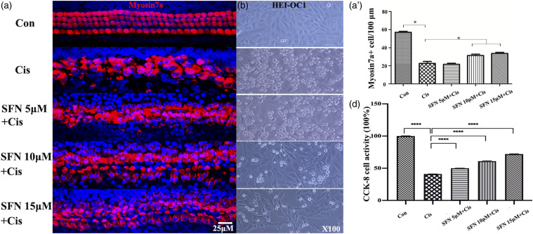
Figure 3.
Sulforaphane protected against cisplatin-induced hair cell damage in vitro. A: Cochlear organotypic culture, with myosin7a staining. A’: Myosin 7a+ cell counting. After cisplatin (10 μM) treatment, the number of myosin 7a+ cells decreased significantly. Myosin 7a+ cell numbers were significantly higher in the sulforaphane +Cis group (10 and 15 μM) comparing to the cisplatin (10 μM) group. B: HEI-OC1 cells culture. Cisplatin (10 μM) induced HEI-OC1 cell death. However, incubated with sulforaphane (5, 10, and 15 μM) prevented cisplatin-induced HEI-OC1 cell death. C: HEI-OC1 cell viability. After treatment with cisplatin (10 μM), HEI-OC1 cell viability decreased to 42%, while sulforaphane (5, 10, and 15 μM) significantly prevented the decrease induced by cisplatin (Scale bar = 25 μm. Red indicates myosin7a, blue indicates DAPI. *p < 0.05, ****p < 0.0001, versus cisplatin group; 3 in each group, n = 15).

