Fig. 2.
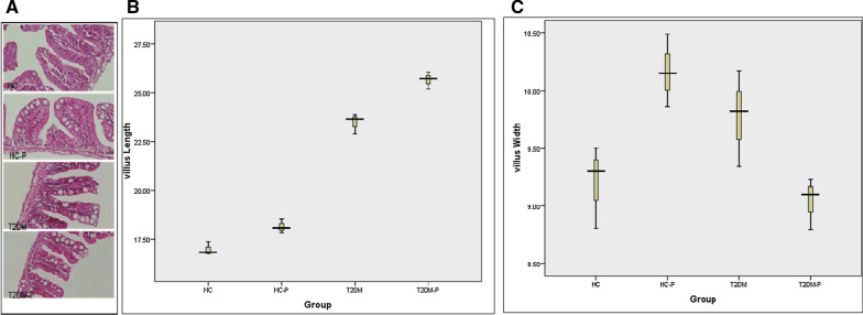
A Histopathology evaluation of zebrafish small intestine. Intestinal tissues were stained with H&E and studied by microscopy (× 400 resolution). Healthy control group (HC), Healthy control group supplemented with probiotics (HC-P), Diabetic group (T2DM), Diabetic group supplemented with probiotic (T2DM-P). B Villus length increased slightly in the T2DM-P group following probiotic supplementation compared to T2DM group. C. Villus width was slightly higher in the T2DM group compared to T2DM-P group. Data shows mean ± SE for three independent assays

