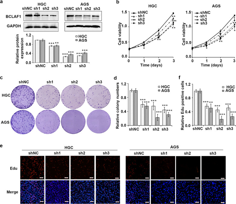
Fig. 4.
BCLAF1 silence inhibits cell proliferation. a Western blot analysis of BCLAF1 expression in HGC and AGS cells infected with shNC or shBCLAF1s (sh1, sh2 and sh3). GAPDH served as an internal reference. b The transfected cells were harvested for MTT assay. BCLAF1 depletion inhibited cell proliferation of HGC and AGS gastric cancer cells. c Colony formation assay was performed to investigate colony formation ability of shNC or shBCLAF1s cells. d Quantitative results of colony formation analyzed with Image J. e EdU assay was used to examine the cell proliferation ability of shBCLAF1s transfected cells. Scale bars, 100 µm. f Three different fields were randomly chosen and quantitative results of EdU assay analyzed with Image J. Data presented as the mean ± SD of three independent experiments. *p < 0.05, **p < 0.01 and ***p < 0.001 vs. shNC based on Student's t-test

