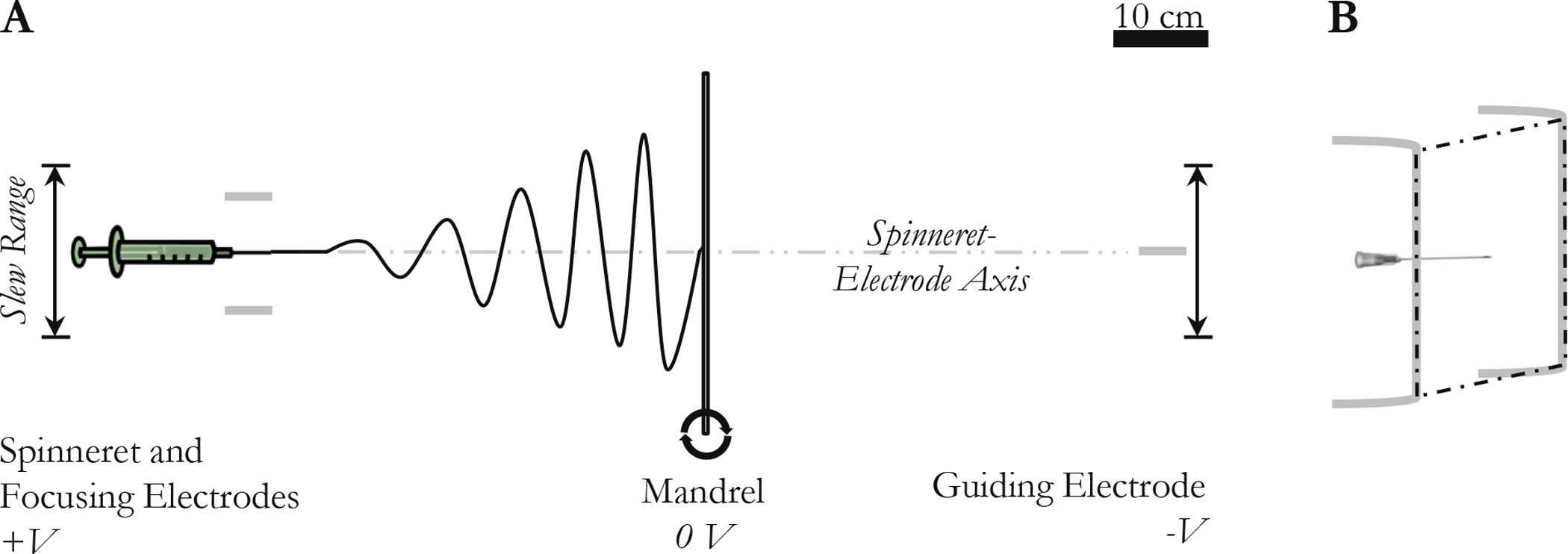
Figure IV–1.

PGS electrospinning schematics. (A) Scale diagram of electrospinning system viewed from the top. The spinneret and the guiding electrode move in parallel so that the spinneret-electrode axis is always perpendicular to the mandrel. (B) The spinneret and focusing electrodes viewed from the top right. The tip of the needle and the vertical portions of the focusing electrodes are coplanar.
