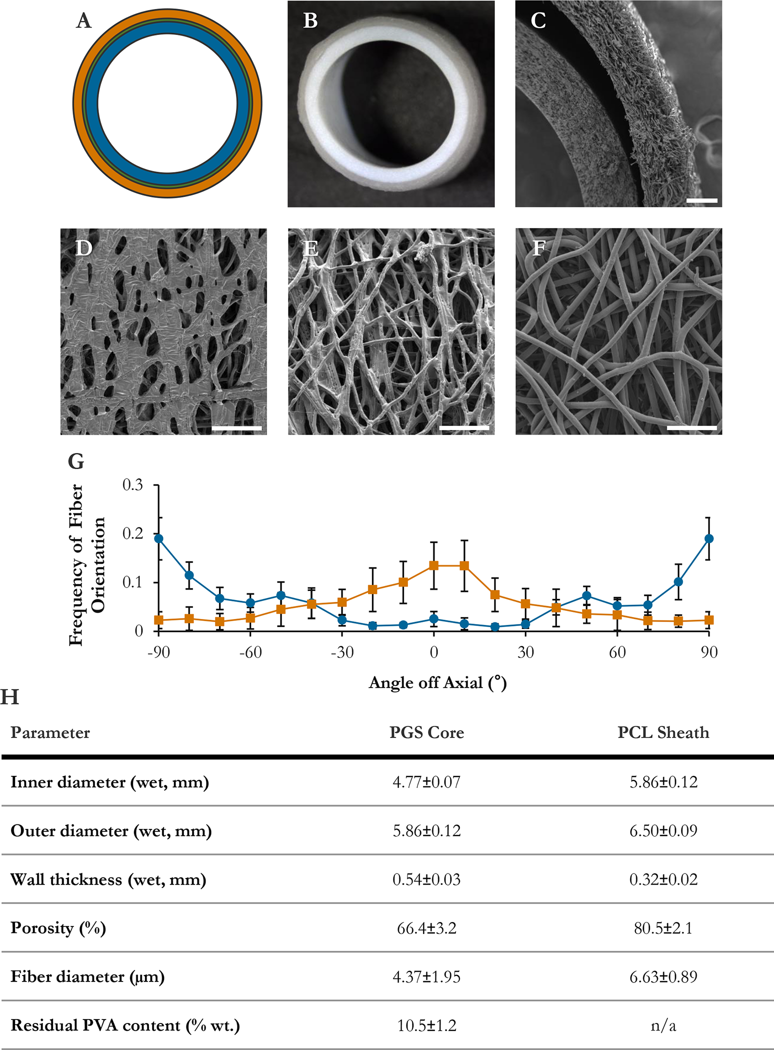
Figure IV–2.

Graft morphology. (A) Scale schematic of graft design. Blue: PGS core. Green: pPGS coating (thin). Orange: PCL sheath. (B) Macroscopic image of wet graft cross-section. (C) Graft wall in transverse. (D) Lumen of PGS core. (E) Ablumen of PGS core. (F) Ablumen of PCL sheath. (G) Fiber orientation distributions in core (blue, circles) and sheath (orange, squares), binned in 10° increments. (H) Graft physical parameters. Bars are 200 μm (C) and 50 μm (D, E, F).
