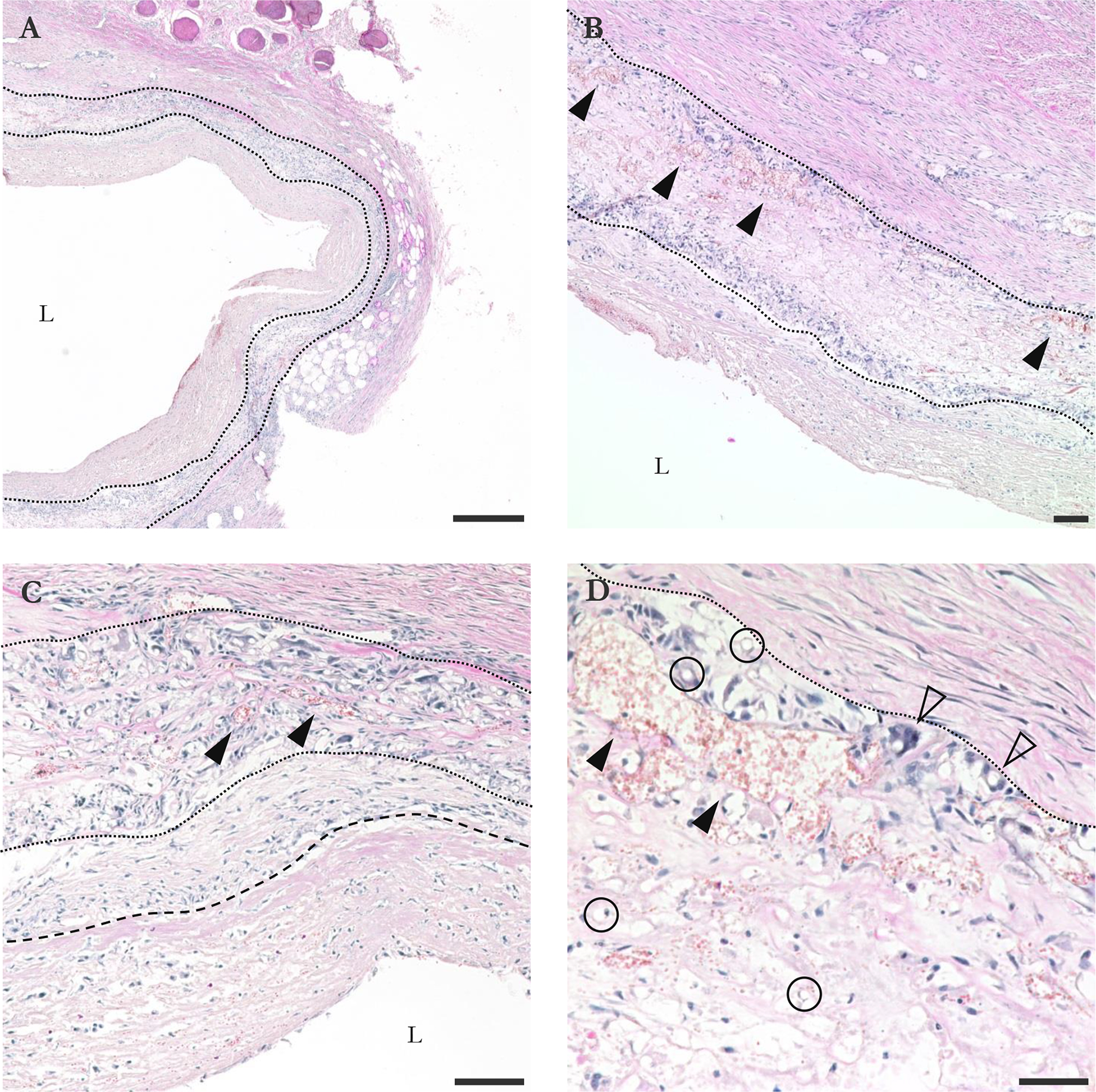
Figure IV–6.

Transverse H&E sections of graft explanted at 15 d. (A) 4X view. Tears in inner and outer capsule are sectioning artifacts; tissue was delicate. Graft material is bounded by dotted lines. (B) 10X view. Solid arrowheads point out some of many capillaries within the graft wall. (C) 20X view. Transition between fibroblast-enriched area and less cellular area of inner capsule marked by dashed line. (D) 40X view of graft-outer capsule interface. Circles show some of many fibers, more easily identified in transverse. Hollow arrowheads show some of many multinucleated monocytes/macrophages. L indicates lumen. Bars are 500 (A), 100 (B, C), and 50 μm (D).
