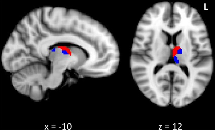
Figure 2.

Thalamic atrophy associated with psychosis and perceptual abnormalities in patients. Legend: Reduced grey matter in the anterior and medial regions of the left thalamus in patients with psychosis‐like experiences (red) and sensory perceptual abnormalities (blue), compared to patients without. Clusters are overlaid on the MNI standard brain and significant at p < 0.05, corrected for multiple comparisons.
