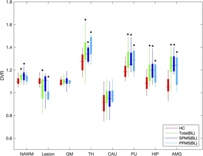
Figure 1.

Comparison with HC (n = 16) and MS baseline (n = 15) in DVR. PK11195 PET DVR among in VOIs in healthy control (HC) as compare to MS patients at baseline scan. Nine secondary progressive (SP) and six primary progressive (PP) patients included. VOIs included normal‐appearing white matter (NAWM), whole lesion mask (Lesion), cortical gray matter (GM), thalamus (TH), caudate (CAU), putamen (PU), hippocampus (HIPP), and amygdala (AMG). *p < 0.05.
