Figure 3.
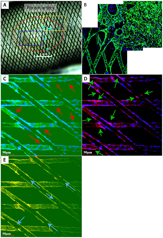
Figure 3 FD implanted aBVM harvested at 14 days. (A) Gross image shows the “aneurysm” neck covered with FD (red circle). Several islands of thin translucent tissue are attached to the struts or filled the pores between struts at the periphery of neck and parent artery interface (arrows). The blue rectangular area demonstrates grossly bare struts at the neck. The whole-mount (B) taken from the yellow rectangular area in panel A shows CD31 positive endothelial cells (green) that are confluent and fully cover the struts in the parent vessel, and are lined along the struts extending to the neck area. (C, D) Taken from the blue rectangular area in panel A, these panels show the “bare struts” are wrapped with a single layer of cells that are not confluent yet and stained positive for both CD31 (arrows in C, green) and aSMA (arrows in D, red). (E) This panel shows panels C–D merged, demonstrating the single layer of cells wrapping around the struts at the neck and also confirming the bare parts of device struts (arrows). (A) Macrophotograph. (B–E) whole tissue mount immunofluorescence (CD31 (green), aSMA (red) and DAPI nuclear counter stain (blue), laser confocal microscopy (original magnification=waterlens x20). aBVM, aneurysm-like blood vessel mimic; aSMA, α-smooth muscle actin; FD, flow diverter.

