
Figure 5.
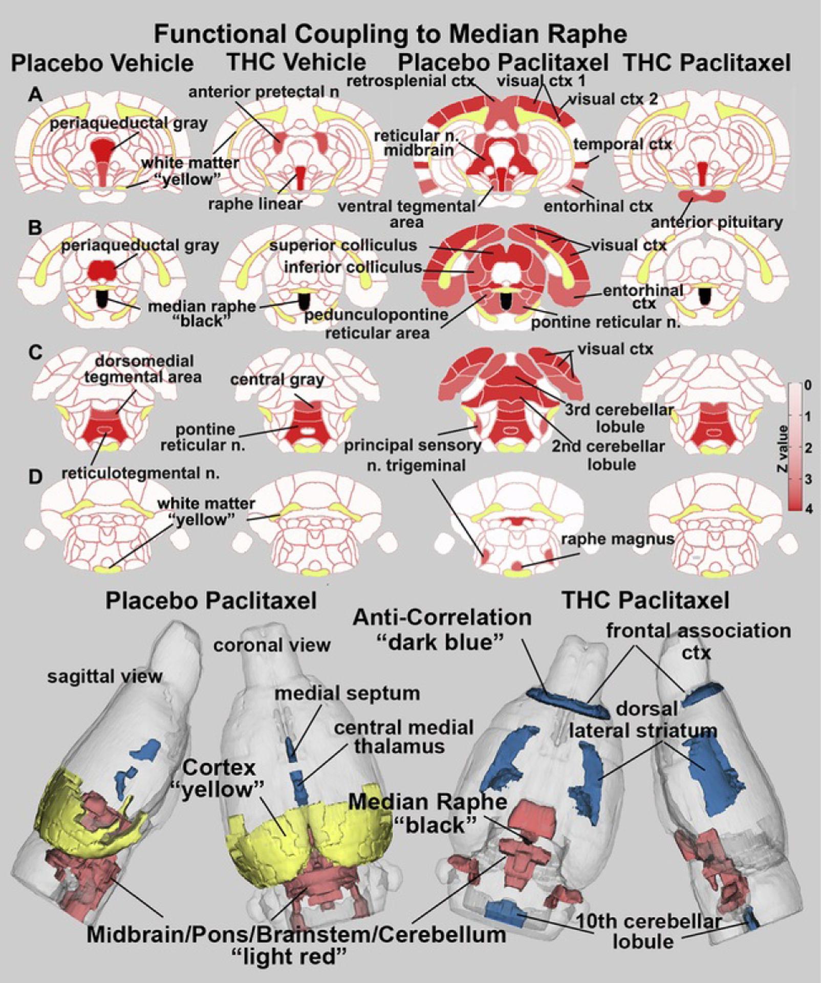
Functional coupling to the median raphe. Paclitaxel produces hyperconnectivity of functional coupling to the median raphe that is attenuated by vaporization of THC-enriched cannabis. The columns of 2D images show heat maps of significant connectivity (z values) for each experimental condition. The shades of red (positive connectivity) appear on anatomical sections taken from the rat brain atlas. These sections are identical across rows (A–D). Yellow highlights white matter tracts, and black shows the location of the median raphe in (B). The brain areas with significant connectivity to the median raphe were taken from Table 2. The most rostral and caudal brain areas showing negative coupling in Table 2 do not appear in the 2D images but are shown in blue in the color-coded 3D reconstruction of placebo paclitaxel and THC paclitaxel conditions below. ctx, cortex; n., nucleus; THC, Δ9-tetrahydrocannabinol.