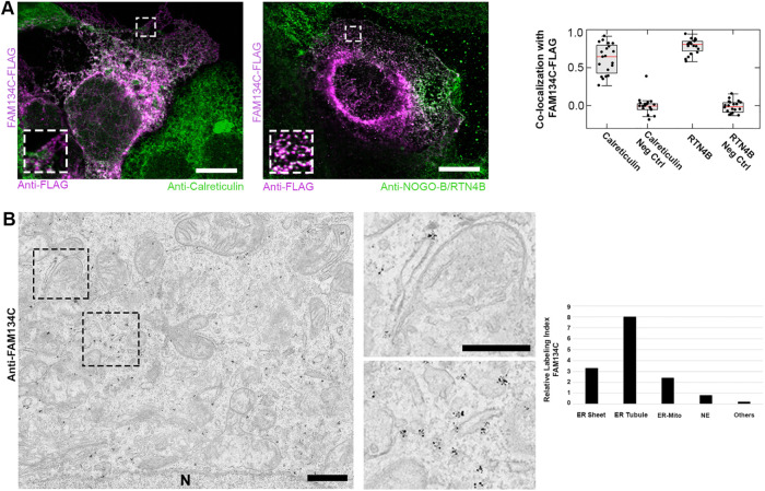
FIGURE 3:
FAM134C localizes to the ER. (A) Confocal LM images of Huh-7 cells overexpressing FAM134C-FLAG (magenta) show colocalization between FAM134C and the general ER marker calreticulin and tubular ER marker RTN4B (green). Insets show higher magnification of boxed areas. Colocalization was quantified using Pearson’s correlation coefficient (∼20 cells). For the negative control (Neg Ctrl) one of the channels from the same ROI was rotated by 90°. Error bars indicate ±SD. (B) Immuno-EM micrographs of U2-OS cells revealed localization of endogenous FAM134C to tubules and sheet edges (insets show higher magnification of boxed area). Scale bars: 10 μm (A), 1 μm (B), and 200 nm (B inset).

