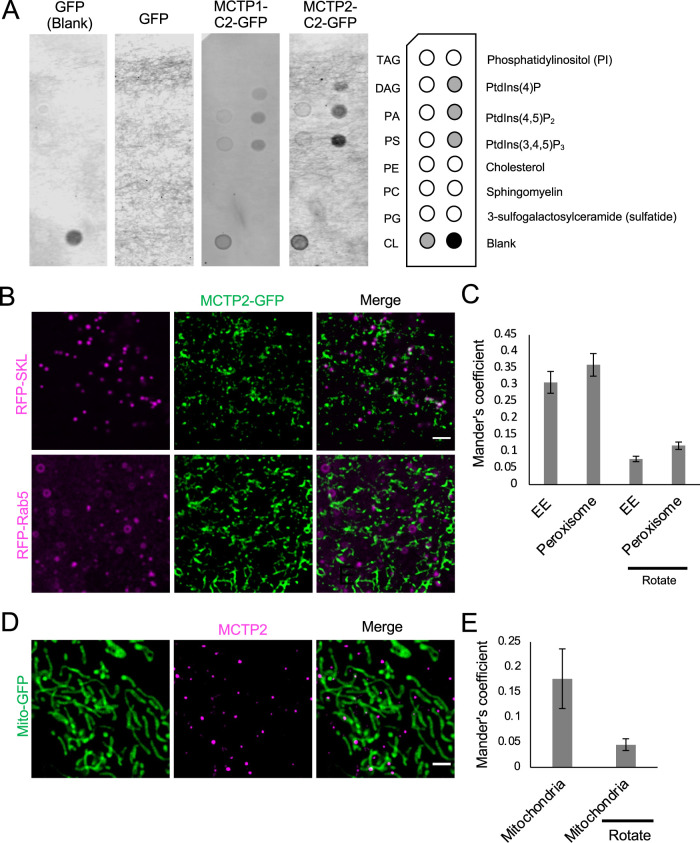
FIGURE 5:
MCTP2 is at ER contact sites with multiple organelles. (A) Lipid binding of C2 domains of MCTPs. List of lipids found on the Echelon lipid strip (on the right). Lysate of cells expressing GFP only or GFP-tagged C2 domains of MCTP1 or MCTP2. Lipid-protein interactions were revealed with anti-GFP antibody. (B) Airyscan images of live cells transfected with mCherry-Rab5 or RFP-SKL and MCTP2-GFP. Scale bar, 2 µm. (C) Quantification of colocalization of mCherry-Rab5 or RFP-SKL and MCTP2-GFP by Mander’s colocalization coefficient. Error bars indicate mean ± SE, n = 22 and 28 fields of view for RFP-SKL and mCherry-Rab5, respectively. (D) Airyscan images of fixed cells transfected with Mito-GFP and stained for MCTP2. Scale bar, 2 µm. (E) Quantification of colocalization of MCTP2 punctae and Mito-GFP by Mander’s colocalization coefficient. Error bars indicate mean ± SE, n = 23 fields of view.

