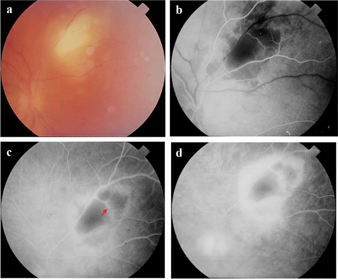
Fig. 6.
Retinal imaging in a patient with ocular toxoplasmosis. a Primary acute toxoplasmic retinitis without other lesions in the surrounding area. b In the arterial phase of fluoroangiography (FA) a masking effect corresponds to the inflamed retina. c In the mid-venous FA phase vasculitis is indicated by a red arrow. d Hyperfluorescence of the inflammatory lesion during the transit FA phase (leakage from the dilated vessels in the area of the lesion). Note that the optic disk is also involved

