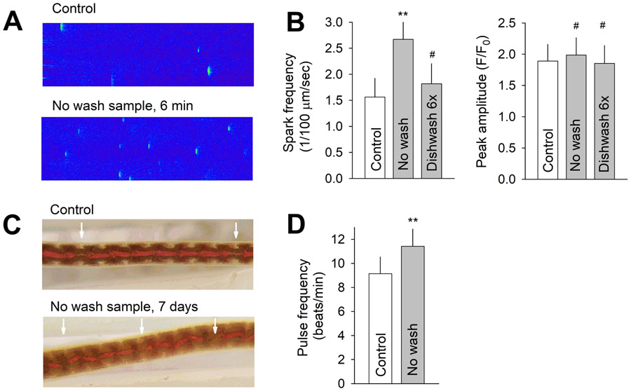
Figure 4.
(A) Representative recordings of Ca2+ sparks in a quiescent mouse ventricular myocyte under BPA-free control and 6 min after exposure to solution prepared using water extract from unwashed tested bottles (No wash). (B) Average Ca2+ spark frequency (left) and peak spark amplitude (right) under control and upon exposure to indicated solutions. N = 15, 8, and 7 myocytes for control, no wash, and dishwashing 6x group, respectively. (C) Representative images of L. variegatus showing the DBV and pulses propagating along the vessel (arrow), after 7-day exposure to control BPA-free solution and solution prepared using water extract from no wash bottles. (D) Average DBV pulsing rate in control and treated groups. N = 21 and 22. ** P < 0.001, # P > 0.1 vs control in unpaired t-test. Error bars are S.D.

