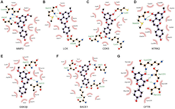
Figure 7.
Molecular docking of GK targets related to Aβ and tau pathology with GK. (A–G) The LigPlus schematic 2D representation of GK-targets interactions. Hydrogen bonds between GK and targets are represented by green dashed lines. The amino acid residues of targets interacted with GK are shown as brown sticks and labeled in green.

