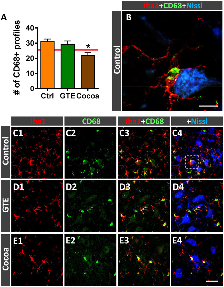
Figure 9.
Impact of GTE- and cocoa-supplemented diets on microglial activation in ventral horn spinal cord of old mice. Sections of lumbar spinal cords from mice of different experimental groups were double immunostained for Iba1 and CD68, a marker of activated phagocytic microglia. (A) Quantification of CD68-positive profiles around MNs in control, GTE and cocoa groups. (B–E4) Representative confocal micrographs used for data analysis showing CD68 (green) in combination with Iba1 (red) and fluorescent Nissl staining (blue, for MN visualization), as indicated in panels. A higher magnification of area delimited by the dashed square in C4 is shown in (B). Data in the graph are expressed as the mean ± SEM; a total of 40-50 images per experimental group were analyzed (number of animals per group: control [Ctrl] = 3, GTE = 4, cocoa = 5). *p < 0.05 vs. Ctrl (one-way ANOVA, Bonferroni's post hoc test). Scale bar: 10 μm in (C) and 50 μm in (E4) (valid for C1–E3).

