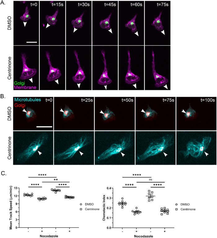
FIGURE 3:
The Golgi appears to nucleate microtubules toward the uropod and nocodazole blocks centrinone effects on the motility of neutrophil-like cells. (A) Time lapse of 500 nM centrinone or DMSO-treated dPLB-985 ManII-GFP cells (labeling the Golgi) migrating on 10 µg/ml fibronectin in an fMLP gradient. Cells were stained with CellMask Orange to visualize the membrane (magenta) and Golgi (green) and imaged every 15 s. White arrowheads mark the leading edge and direction of cell migration. Scale bar: 15 µm. (B) Time lapse of representative DMSO- and centrinone-treated cells depicting microtubule convergence at the Golgi. dPLB-985 cells were stained with 1 µM SiR-Tubulin (cyan) for 1 h and then imaged every 5 s to visualize the Golgi (red). White arrowheads indicate the point of microtubule convergence. Cell images are displayed at 25-s intervals. Scale bar: 15 µm. (C) Mean track speed and chemotactic index of nocodazole-treated (10 µM for 1 h before tracking). dPLB-985 cells ± centrinone (data displayed as mean + SEM; N = 3 repeats, n = 9 devices, 3256–4772 cells). Cells were imaged in a 10-µg/ml fibronectin-coated microfluidic device migrating toward an fMLP gradient every 30 s for 45 min. Significance was determined by mixed-effects REML regression with a Satterthwaite degrees of freedom approximation. n.s. = not significant; **, p < 0.01; ****, p < 0.0001.

