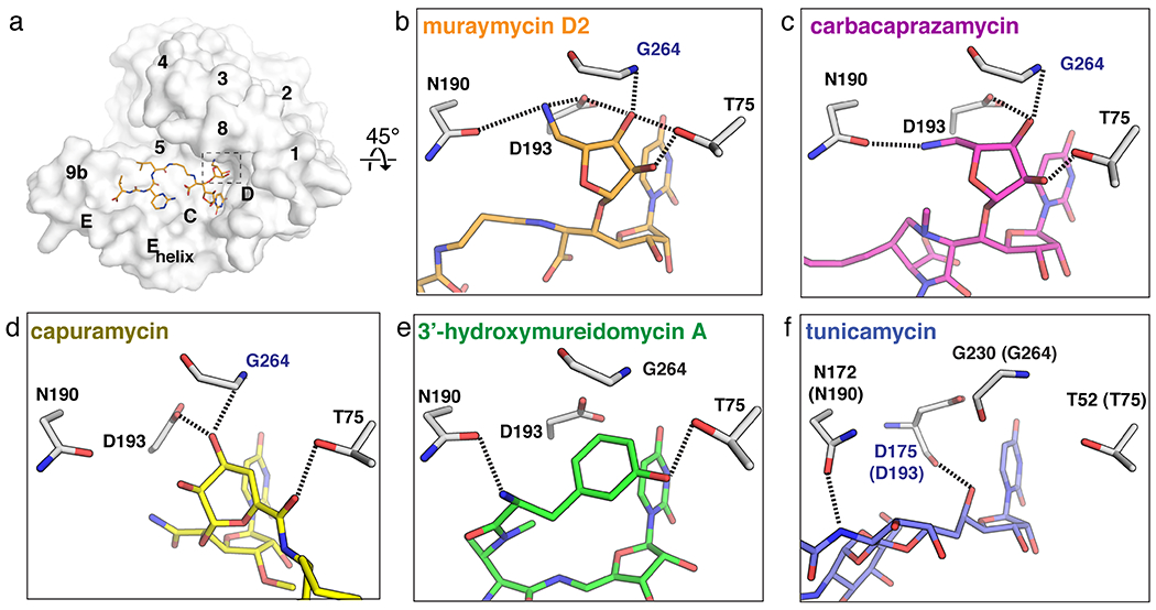
Figure 4.

The uridine-adjacent pocket in MraY. (a) Surface representation of the MraYAA protomer bound to muraymycin D2 (cytoplasmic side). The uridine-adjacent binding pocket is hightlighted with dashed lines. (b)-(e) The uridine-adjacent pocket in each inhibitor-bound structure, oriented 45° relative to the structure in (a). Residue numbering is for MraYAA, except for in panel (f), which shows numbering for MraYCB with numbering for MraYAA in brackets. Residues labelled in blue form backbone interactions with the ligand. Muraymycin D2 PDB ID: 5CKR (orange); tunicamycin PDB ID: 5JNQ (blue); carbacaprazamycin PDB ID: 6OYH (magenta); capuramycin PDB ID: 6OYZ (yellow); 3’-hydroxymureidomycin A PDB ID: 6OZ6 (green).
