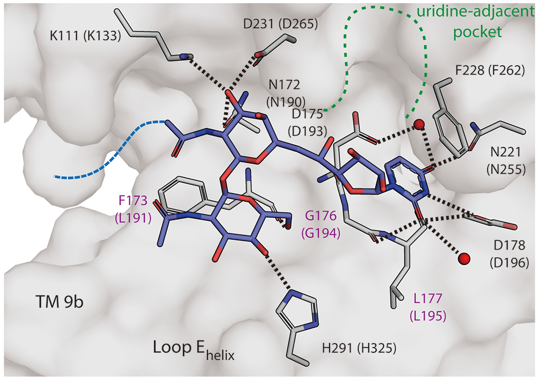
Figure 5.

Detailed view of tunicamycin binding in MraY. Zoomed-in view of the tunicamycin binding site in MraYCB (PDB ID: 5JNQ). The residues forming interactions with tunicamycin (blue) are shown in stick representation (numbering for MraYCB, with numbering for MraYAA in brackets). Residues labelled in purple are forming backbone interactions with tunicamyin. Waters are shown as red spheres. The uridine-adjacent site is delineated with dashed green lines. The aliphatic tail of tunicamycin was disordered and is represented by a dashed blue line.
