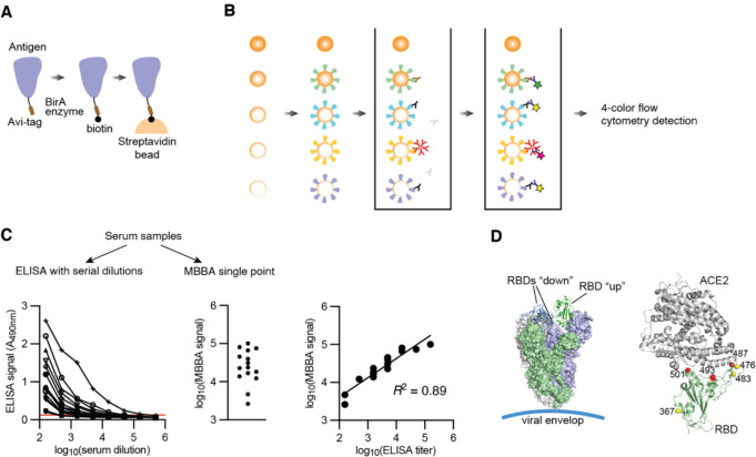
Figure 1.
Design of 2D-MBBA for SARS-CoV-2 antibody profiling.
(A) Scheme showing the design of an Avi-tagged antigen and site-specific immobilization on a streptavidin-coated bead.
(B) Scheme showing the 2D-MBBA principle. Five types of microbeads each presenting a different antigen are mixed and reacted with a serum sample. After washing, bound antibodies are detected with isotype-specific secondary antibodies on a four-color flow cytometry that separately quantify the beads and three isotypes.
(C) Comparison of MBBA with conventional ELISA using serum samples and RBD. ELISA end point titers were determined using the cutoff shown as the red horizontal line. The R2 values were determined after log10 transformation of the ELISA endpoint titers and MBBA signals.
(D) Antigen design. Schematic drawings of the spike protein (PDB ID: 6VSB) and of RBD in complex with ACE2 (PDB ID: 6VW1) denoting mutations used in this study.

