Figure 3.
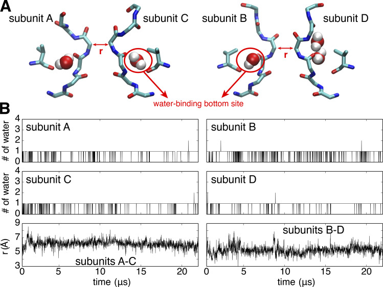
MD simulation of the Shaker channel with an opening of 32 Å for the intracellular gate yields a stable constricted conformation of the selectivity filter. The four channel subunits are labeled A, B, C, and D in clockwise order seen from the extracellular side. (A) Representative conformation of the selectivity filter and water molecules bound behind the filter. (B) Time series of the number of water molecules occupying the water-binding bottom site within each subunit (upper and middle) and the cross-subunit distance between the Cα atoms of G444 of diagonally opposed subunits A and C (lower left) and B and D (lower right) during the 22-µs simulations in the wide-open channel.

