Figure 1.
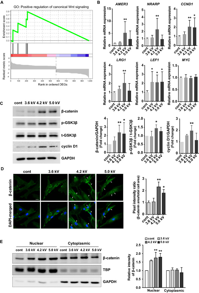
NTAPP activated the Wnt/β-catenin signaling in human dermal papilla (hDP) cells. hDP cells were exposed with NTAPP at 4.2 kV (for RNA-sequencing) or at different plasma doses (3.6 kV, 4.2 kV, and 5.0 kV; for Western blot) for 1 min and were further incubated for 24 h. (A) Gene set enrichment analysis for a gene ontology (GO) gene set, GO:0090263. Normalized enrichment score (NES) = 1.812; nominal p value = 0.014. A total 983 differentially expressed genes (DEGs) was pre-ranked. (B) Quantitative RT-PCR analysis of the expression of selected genes related to the Wnt-β-catenin signaling pathway. Results are expressed as mean ± S.D. of three independent experiments. (C) Western blots on β-catenin, GSK3β, and cyclin D1 in hDP cells after NTAPP treatment at different output voltages (3.6, 4.2, and 5.0 kV). (D) Immunofluorescent staining of β-catenin in hDP cells treated with NTAPP. Staining intensities of nuclear β-catenin staining were quantified using Image J. Arrows highlight nuclear staining. Scale bar = 20 μm. (E) The amounts of β-catenin in both cytoplasmic and nuclear fractions were separately measured. GAPDH and TATA binding protein (TBP) were used for loading controls for cytoplasmic and nuclear fractions, respectively. Results are expressed as mean ± S.D. of three independent experiments. p values were determined by one-way ANOVA. *p < 0.05, **p < 0.01 (vs. control). The original images of blots for (C, E) are available in Supplementary Information.

