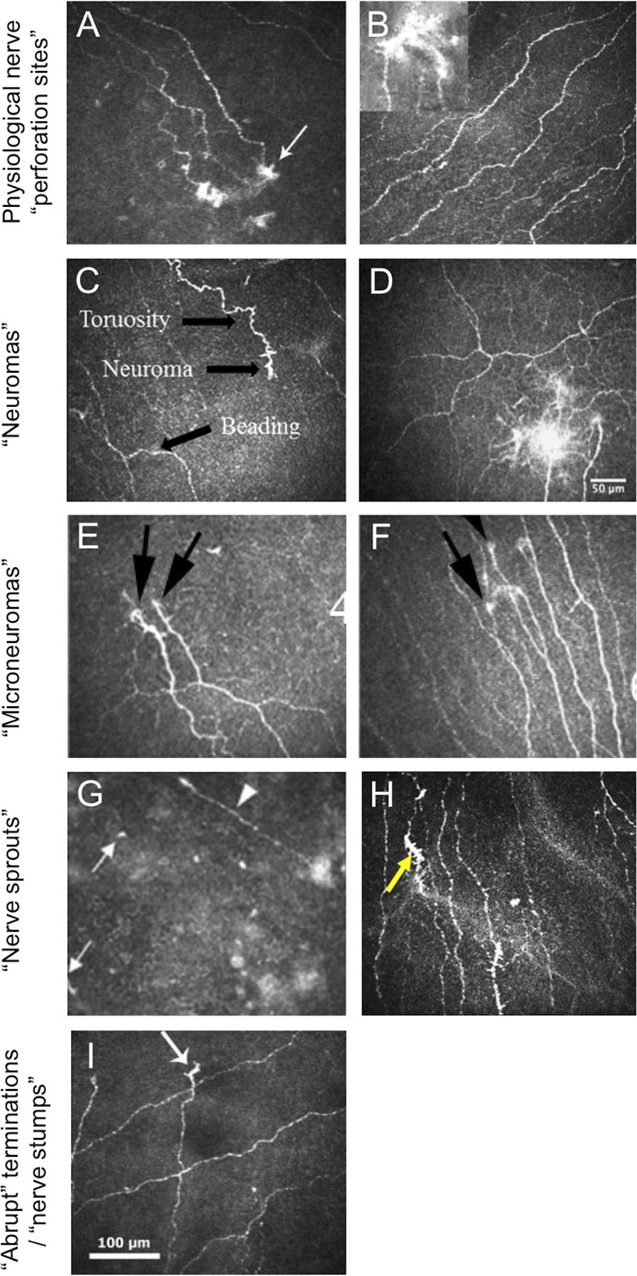
Figure 1. Laser-scanning IVCM images of corneal nerve features, reproduced with permission from papers included in the review.

A. From Patel and McGhee[13] showing “probable sites of perforation of nerves through Bowman’s layer (white arrow) in the infero-temporal mid-periphery”. B. From Al-Aqaba et al.[21] showing “Normal appearance of the sub-basal nerve plexus seen in a healthy control. Bulb-like termination of sub-basal nerves is shown in the inset.” C. From Aggarwal et al.[11] showing a “neuroma” from a patient with neuropathy-induced severe photoallodynia. D. From Cruzat et al.[2] of “multiple neuromas” in a patient with corneal allodynia. E. and F. Both from Dieckmann (2017)[34] from individuals with neuropathic corneal pain showing “presence of micro-neuromas (black arrows)”. G. From Lagali et al.[27] identifying “presumed sprouting subbasal nerves (white arrows) and a regenerating subbasal nerve (arrowhead)” after phototherapeutic keratectomy. H. From Shen et al.[36] showing “nerve sprouts” (yellow arrow) in an individual with episodic migraine. I. From Patel et al.[22] showing “apparent abrupt terminations (arrow) of sub-basal nerve fiber bundles within the region of the cone in severe keratoconus.
