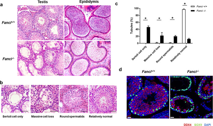
Fig. 3. FANCI deletion causes germ cell loss in mice.
a H&E staining of testes and epididymides from 8 weeks old wild type and Fanci−/− mice. Scale bars, 50 μm. b Representative images of H&E stained testicular sections showing various seminiferous tubules in 8 weeks old Fanci−/− mice, including Sertoli cell-only tubules, tubules with massive cell loss, tubules with round spermatids as the most advanced spermatogenic cells, and relatively normal tubules. Scale bars, 50 μm. c Quantification of different types of seminiferous tubules in 8 weeks old wild type and Fanci−/− mice. Six wild-type mice and six Fanci−/− mice were analyzed. Data are presented as mean ± SD. *, P < 0.05. Chi-square test (Fisher’s exact test). d Immunofluorescence staining for DDX4 (a germ cell marker) and SOX9 (a Sertoli cell marker) in wild type and Fanci−/− mice testes of 8-week-old mice. Scale bars, 20 μm.

