Fig. 1.
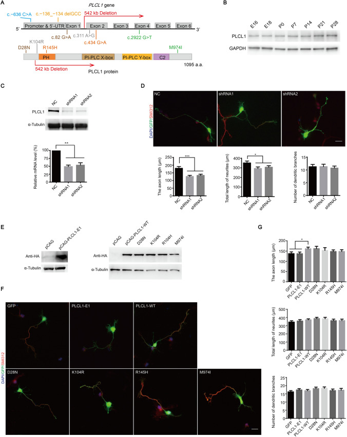
The dosage of PLCL1 is crucial for neurite outgrowth of primary cultured mouse cortical neurons, and the deletion of PLCL1 exons 2–6 affects the promotion of neuronal growth, while PLCL1 missense variants have no effect. A The location of PLCL1 rare variants in studied ASD patients on the PLCL1 gene and coding protein. Colored bars represent the PLCL1 protein functional domains. B The PLCL1 expression pattern in mouse cerebral cortex during the embryonic and postnatal period. C The efficiency of endogenous Plcl1 knockdown was verified using shRNA packaged in lentivirus by Western blot (top) or using pFUGW-H1-shRNA plasmids by qRT-PCR (bottom) in primary cultured mouse cortical neurons. D Plcl1 knockdown affects mouse neuronal growth. Upper panel shows the specific cellular morphology of neurons transfected with pFUGW-H1 vector (NC, as a control), Plcl1 shRNA1 and shRNA2. All neurons were co-labeled with DAPI (to identify nuclei), GFP (to identify overall neuronal morphology) and SMI 312 (an axonal marker). Scale bar, 20 μm. Bottom panel shows the statistical results of the axon length, total length of neurites, and the number of dendritic branches of mouse neurons with Plcl1 knockdown. Approximately 90 cells from three independent experiments were counted and the P-value was determined by one-way ANOVA. *P <0.05, **P <0.01, ***P <0.001. E Western blot analysis of ectopic expression of PLCL1-WT, PLCL1-E1, and four missense variants in HEK-293T cells. F The morphology of neurons overexpressing the CAG vector (GFP, as a control), PLCL1-WT, PLCL1-E1, and four missense variants. Scale bar, 20 μm. G The axon length (upper), total length of neurites (middle), and the number of dendritic branches (bottom) were measured and analyzed as described above. Error bars, ± SEM.

