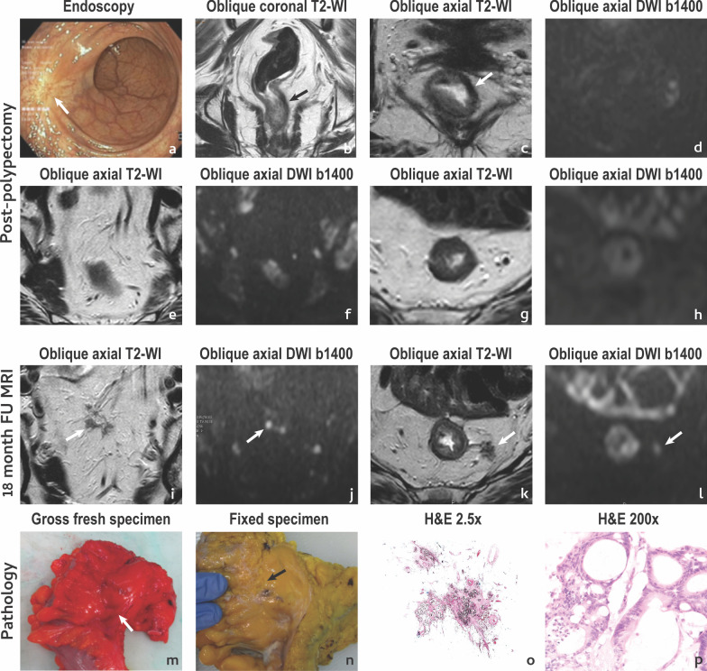
Fig. 10.
Extra-rectal pelvic recurrence. A 62 year-old female with rectal bleeding presented with a 34 mm polyp at colonoscopy which was excised revealing a tubulovillous adenoma with moderately differentiated adenocarcinoma, mucinous type, invading the muscularis propria with a focally positive margin in depth. No residual tumour was apparent on MR imaging or endoscopy at post-polypectomy assessment (a–d) and no extra-rectal suspicious findings were found either (e–h). Patient underwent long course chemoradiation and was followed. Findings were stable until the 18th month of follow-up, when irregular, heterogeneous, intermediate signal foci were found cranially to the polypectomy scar, within the mesorectal fat, at two different levels (arrows in i, j and k, l). Patient underwent total mesorectal excision and the MR imaging findings corresponded to extranodal tumour deposits of intestinal type adenocarcinoma with mucinous areas. The extranodal tumour deposits in i, j are shown in the fresh (m) and fixed (n) pathology specimen and at hematoxilin eosin staining both at low (× 2.5) (o) and high (× 200) (p) magnification

