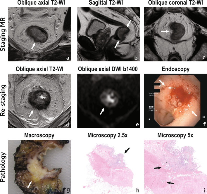
Fig. 2.
Incomplete/poor tumour response. A 75 year-old male presented with a low mrT3a mrN0 EMVI + (not shown) CRM-rectal cancer (arrows in a–c). He underwent NAT and was re-staged at 12 weeks with MR imaging (d, e) and endoscopy (f). Reduction in size was estimated as < 50%, as may be inferred in d vs a. On T2-WI, the tumour scar was composed largely of intermediate signal intensity tissue (d), classified as mrTRG4 with absent split scar sign. On high b value DWI (e), a thick layer of high signal intensity was apparent at the endoluminal aspect of the tumour bed and a persistent infiltrative lesion was visible on endoscopy (between arrows in f). Patient underwent surgery and specimen was staged as a ypT3 (extension into mesorectal fat visible at macroscopy in g) N1c (not shown) TRG3 R0. At microscopy, viable tumour was predominantly mucosal/submucosal (arrow in h) but there were niches of viable tumour cells within the muscularis propria (arrows in i) and also at perirectal fat (not shown)

