Fig. 6.
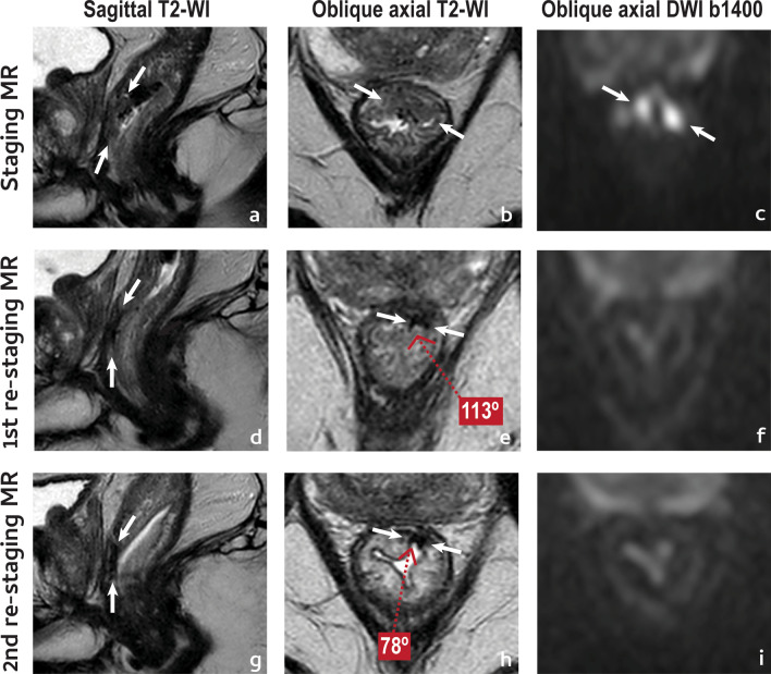
Complete tumour response. A 61 year-old male presented with a low anterior mrT2 (between arrows in a–c) mrN1a (not shown) EMVI− rectal cancer. He underwent NAT and was re-staged at 11 weeks (d–f). Reduction in tumour size was considerable, > 80% (d, e). On T2-WI, the tumour was reduced to a thin, crescentic, endoluminal hypointense “fibrotic” scar (mrTRG1, split scar sign+) (e). On high b value DWI, no high signal intensity was visible (f). Notice how the scar “curled-in” between first and second assessment (e, h, respectively), reducing its depth angle significantly (− 35°). Patient is currently on W&W with no signs of disease recurrence at 1 year

