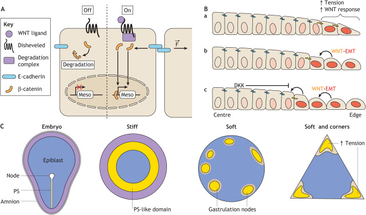
Fig. 5.
Convergence of WNT signalling and mechanics. (A) Diagram illustrating how both canonical WNT signalling and tension at cellular junctions regulate the intracellular level of active β-catenin. Once in the nucleus, β-catenin activates mesodermal genes (Meso). F indicates force applied at the cellular junction. (B) Mechanism of patterning in WNT-treated gastruloids. Although WNT is provided uniformly, cell responsiveness is highest at the periphery, where the cells fail to form a mature epithelium (a). WNT positively regulates its expression at the same time as promoting epithelial-to-mesenchymal transition (EMT) (downregulating E-cadherin) (b). As secreted WNT diffuses, it activates the pathway in neighbouring cells. Concomitantly, EMT in the first cell destabilises junctional β-catenin in neighbouring cells. This phenomenon propagates from cell to cell in an inward direction until a sufficiently high level of secreted WNT inhibitor (DKK) terminates the process (c). (C) Comparative diagram of the patterning in embryos and BMP4-treated human PSC colonies, whether these colonies are micropatterned on stiff plastic dishes or soft hydrogels.

