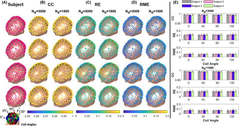
Fig. 8.
(A) A total of 256 coil positions were defined around the scalp surface of each subject. The inset shows the four coil orientations used. (B-D) The spatial profiles of the error metrics CC, RE and RME averaged across the four coil orientations at each location for both 3000 and 1500 dipole basis function sets. (E) The mean and std of the error metrics shown across subjects and coil orientations.

