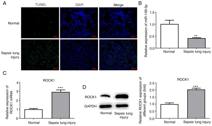
Figure 3.
Effects of ALI induction on cell apoptosis, and the expression of miR-139-5p and ROCK1. (A) Histological analysis of mouse lung tissue samples using TUNEL staining. Representative histological images (magnification, x400) and percentage of positively stained cells. Scale bar, 100 µm. (B) miR-139-5p levels in lung tissues were assessed using RT-qPCR analysis. ROCK1 levels in lung tissues were assessed by (C) RT-qPCR and (D) western blotting. Data are presented as the mean ± standard deviation. **P<0.01, ***P<0.001 vs. normal group. RT-qPCR, reverse transcription-quantitative PCR; ALI, acute lung injury; ROCK1, Rho-associated kinase 1; miR, microRNA.

