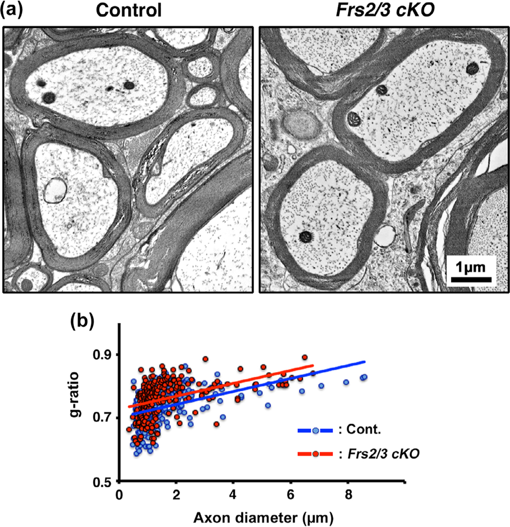
FIGURE 5.
Complete ablation of Frs2/3 in oligodendrocytes did not lead to a significant reduction in myelin thickness in the spinal cords of adult mice. (a) EM images taken from matched regions of the ventral cervical spinal cord white matter at 5 months of age show that the thickness of myelin in the Frs2/3 cKO mice appears to be similar to that in their littermate controls. (b) Quantification of myelin thickness by g-ratio analysis, presented as scatter plots relative to axon diameters shows a trend towards thinner myelin sheaths in the mutants, indicated by slightly higher g-ratios in the Frs2/3 cKO mice (red dots; average g-ratios = 0.762 +/−0.004) compared to its littermate control (blue dots; average g-ratios = 0.736 +/− 0.004, p = 9.35 × 10⁻⁷). Approximately 200–400 axons from two mice of each genotype were analyzed. Representative images of spinal cords taken from similar regions of lateral-ventral white matter from the control and mutant mice are shown. Scale bar, as indicated [Color figure can be viewed at wileyonlinelibrary.com]]

