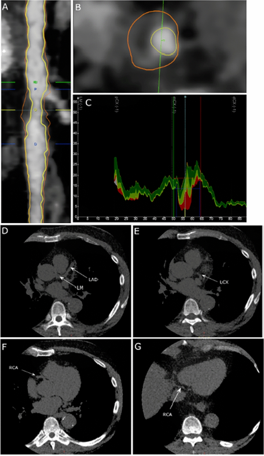
Fig. 1.

Plaque analysis with semiautomatic plaque analysis software. A Longitudinal straightened multiplanar reconstruction of left circumflex artery featuring a high-risk plaque (HRP) between the blue lines. B Transverse vessel view demonstrating HRP with positive remodeling, napkin ring sign, and low attenuation core (< 30 HU). C Graph depicting lumen and vessel areas as a function of vessel length. Plaque subtypes dense calcium, fibrous, fibrous-fatty and necrotic core are shown in grey, green, light green, and red colors. The bottom panels shows unenhanced axial images in the same patient demonstrating no coronary artery calcium present in the left main (LM) and left descending artery (LAD) (D), left circumflex artery (LCX) (E), and right coronary artery (RCA) (F, G)
